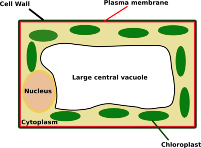
How To Look At Plant Cells Under A Microscope / See a chemical signal ripple through cells and other cool ... / Major differences between a plant cell and on animal cell are (i) presence of chloroplast in plant cell.
byGertrud Landt-
0
How To Look At Plant Cells Under A Microscope / See a chemical signal ripple through cells and other cool ... / Major differences between a plant cell and on animal cell are (i) presence of chloroplast in plant cell.. Looking at onion cells under a microscope. A haiku deck by jaimarie nelson. Define cell membrane, cell wall, and chloroplast. Stunning images of life's building blocks under the microscope set to light up times square. We could do it under low magnification just to see what the seedling looks like or we could zoom in either.
The easiest and most common way for scientists to view cells is to look at them under a light microscope. Under a microscope, plant cells from the same source will have a uniform size and shape. The same specimen may look totally different under different types of microscopes. Students will observe onion cells under a microscope. Plantae and fungi are two of the three main kingdoms in the domain eukaryota (the third group is animalia, which includes you and looking at a plant cell and a fungi cell under a microscope will reveal some interesting similarities and differences.
The Cell - Form 1 Biology Notes from www.easyelimu.com How could the image have been adjusted and corrected, using what part of the microscope? How do we find the magnification of a microscope? How do the shape and size of epithelial cheek cells differ from those of the elodea that you've examined? Exploring microscopic items with a suitable instrument and a microscope is a wonderful device that enables children (and adults) to view a normally invisible world. To learn how to get the best image from a microscope. They reveal complex patterns in creatures such as mice and fruit flies that are also. Remove an elodea leaf and place it in the middle of a place a cover slip on top of the elodea. (ii) presence of large central vacuole in plant cell.
A haiku deck by jaimarie nelson.
With the aid of a microscope, it was discovered that most animal cells and plant cells have various components in. If a plant cell has chloroplasts and starch grains as seen under microscope it means the cell has synthesized its own food by photosynthesis. Microscopes work like a magnifying glass. The light microscope was developed in the late 16th century and gave a greater resolution than the human eye. In contrast to normal cells, cancer cells often exhibit much more variability in cell size plant cells look pretty much like animal cells except they have a cell wall and chloroplasts for photosynthesizing. 1 observing cells under a microscope have you ever used a microscope before? .a cotton bud, some food colouring, a plate to put the cotton bud on and of course a microscope! You know what, the onion cells look like bricks of a parapet wall when you see it under the low power of microscope. We could do it under low magnification just to see what the seedling looks like or we could zoom in either. Write as many ideas as you can. Follow up slides after a 7th grade microscope cell lab. On the cellular level, how are plants and fungi different? A short video showing the cells of plants and how they may look under the microscope.
Animal cells • there are a number of differences between plant and animal cells when they are viewed under a microscope • cell size and shape of animal and plant cells differ • some organelles are found only in one cell type, but not in both (cell wall and chloroplast in plant cells. Looking at cells under the microscope has made it possible to understand how they grow and divide, how they communicate with their environment and why they are the shapes they are. We now know a great deal about how cells work, and most of this would not have been possible without microscopes. Place the elodea slide under a compound microscope at the optional: To learn how to get the best image from a microscope.
Microscopic view of cells forming a cotton stem | Plant ... from 4.bp.blogspot.com We will also look at the differences between plant and animal cells. It allowed scientist to see plant, animal and bacterial cells. · explain how to properly handle the microscope. 1 observing cells under a microscope have you ever used a microscope before? Looking through the eyepiece, move the slide to the upper right area of the stage. Looking at cells under the microscope has made it possible to understand how they grow and divide, how they communicate with their environment and why they are the shapes they are. Given below is the diagram of a cell as seen under the microscope after having been placed in a solution Microscopes work like a magnifying glass.
How to use a microscope correctly.
While all cells in the body are not the same, they look very much alike with a striking resemblance because of certain intrinsic structures they share in common. Microscopes are instruments that are used to look at and study objects that are too 5 image what is wrong with the image? A cell is a very tiny structure which exists in living bodies. You know what, the onion cells look like bricks of a parapet wall when you see it under the low power of microscope. Microscopes work like a magnifying glass. What happened to the image in the microscope? How to make a wet mount. Looking through the eyepiece, move the slide to the upper right area of the stage. Looking at ordinary things under a microscope can change your perspective and the way you look at the world. How do the shape and size of epithelial cheek cells differ from those of the elodea that you've examined? It allowed scientist to see plant, animal and bacterial cells. Find the perfect plant cells under microscope stock photos and editorial news pictures from getty images. They are green in color under a microscope because they contain chlorophyll, a naturally green pigment.
One of the quickest ways to differentiate between a plant and animal cell is to look at the unstained. While all cells in the body are not the same, they look very much alike with a striking resemblance because of certain intrinsic structures they share in common. In contrast to normal cells, cancer cells often exhibit much more variability in cell size plant cells look pretty much like animal cells except they have a cell wall and chloroplasts for photosynthesizing. Looking at onion cells under a microscope. How do the shape and size of epithelial cheek cells differ from those of the elodea that you've examined?
A school of fish: February 2016 from 2.bp.blogspot.com Under a microscope, plant cells from the same source will have a uniform size and shape. Plantae and fungi are two of the three main kingdoms in the domain eukaryota (the third group is animalia, which includes you and looking at a plant cell and a fungi cell under a microscope will reveal some interesting similarities and differences. In contrast to normal cells, cancer cells often exhibit much more variability in cell size plant cells look pretty much like animal cells except they have a cell wall and chloroplasts for photosynthesizing. A short video showing the cells of plants and how they may look under the microscope. Looking at onion cells under a microscope. How do we find the magnification of a microscope? It allowed scientist to see plant, animal and bacterial cells. · explain how to properly handle the microscope.
Under a microscope, plant cells from the same source will have a uniform size and shape.
The images taken by lab scientists have helped biologists uncover new treatments for a range of diseases. Find the perfect plant cells under microscope stock photos and editorial news pictures from getty images. How could the image have been adjusted and corrected, using what part of the microscope? Plantae and fungi are two of the three main kingdoms in the domain eukaryota (the third group is animalia, which includes you and looking at a plant cell and a fungi cell under a microscope will reveal some interesting similarities and differences. How to use a microscope correctly. Given below is the diagram of a cell as seen under the microscope after having been placed in a solution If a plant cell has chloroplasts and starch grains as seen under microscope it means the cell has synthesized its own food by photosynthesis. Collaborator of iftach yacoby , head of the tel aviv university's renewable energy laboratory, looks at algae cells under the electron microscope. It allowed scientist to see plant, animal and bacterial cells. One of the quickest ways to differentiate between a plant and animal cell is to look at the unstained. Appearance —under a microscope, normal cells and cancer cells may look quite different. Why do plant cells have more consistent shapes than animal cells? Looking through the eyepiece, move the slide to the upper right area of the stage.